Thousands of shades of grey - on the UBC MRI Research Centre
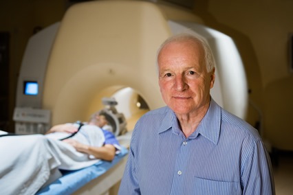 The Canadian Olympic swim team has been inside it. Compulsive gamblers have been inside it. Mostly, however, patients have been inside it – people with a range of afflictions, including Parkinson’s disease, schizophrenia, and multiple sclerosis.
The “it” is a hulking, humming, clanking machine in the basement of UBC Hospital – a 3-Tesla magnetic resonance imaging scanner, or MRI, that is the centerpiece of the UBC MRI Research Centre.
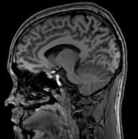
This year, the centre marks its 10th year, and it’s busier than ever, with the scanner booked for a wide range of research projects – especially those focused on the brain, because it provides detailed images of soft tissue that are impossible with X-rays or computed tomography, and are easier and safer than positron emitted tomography (PET).
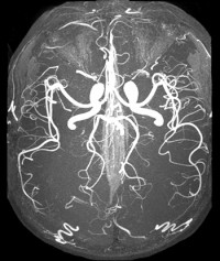
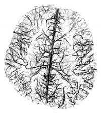
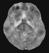
The machine is sought after because it’s not your ordinary MRI. A 3-Tesla scanner is twice as strong as clinical MRIs, producing a magnetic field 60,000 times stronger than Earth’s magnetic field. When used with contrast agents, the images can contain over 100,000 shades of grey. It is one of only two in B.C.; the other was installed last year at BC Children’s Hospital.
Playing with physics and mathematics
 But even as researchers exploit the machine for their own projects, the MRI Research Centre also has found ways to tease even more revealing data from the technology.
“MRI scanners are relatively open hardware platforms, similar to smartphones,” said Alex Rauscher, a physicist at the centre and an Assistant Professor in the Department of Radiology. “Just as a smartphone comes alive due to its apps, so does the MRI scanner. We are able to play with the physics and mathematics behind these things and understand what tissue changes do to the MRI signal.
If such a technique is proven to be successful, it becomes part of the portfolio of scans included by the manufacturer in their newer machines.
Dr. Rauscher, for example, has developed a sensitive method of improving image resolution by increasing the ratio of “signal” (useful information) to “noise” (background or irrelevant information). The signal-to-noise ratio is so high that it yields more than just stunningly clear images; it also provides precise numbers that delineate the structure of tissue, such as lesions too small to be visually discerned, and the concentrations of various biochemicals.
“You can be a chemist, measuring the outcomes of reactions,” said Alex MacKay, the founding Director of the MRI Research Centre, and a Professor in the Department of Radiology and the Department of Physics and Astronomy. “But to do that, you have to understand the physical properties of the molecules you’re measuring. And you have to understand the signal when the hydrogen atoms in your body are ‘flipped’ by the scanner’s magnetic field. So you also have to be a physicist – or at least have one at your side.” (Three of the centre’s faculty members are physicists, including Dr. MacKay and Dr. Rauscher.)
Measuring myelin
One of the most fruitful targets for the MRI Research Centre has been myelin, the electrical insulation layer surrounding neurons. Multiple sclerosis (MS) results from a breakdown of myelin – when it wears away, the brain’s electrical signaling slows down.
Dr. MacKay has developed a way of capturing images of myelin in living humans, by homing in on the water inside it. That technique has helped make MRI a valuable tool for understanding MS’s progression. Refinements of the technique are enabling MS researchers, such as Tony Traboulsee, an Associate Professor of Neurology and Medical Director of the UBC Hospital MS Clinic, to determine if potential therapies can slow down de-myelination, or stimulate re-myelination.
 The centre was created with a grant from the Canada Foundation for Innovation, which funded the 2003 purchase of the 3T scanner – at the time, an almost experimental machine, made by Philips, the Dutch multinational electronics and engineering company. The centre later acquired another, even stronger MRI, a 7T, but it is much smaller, used mostly for scanning animal models of various diseases.
The centre’s 3T machine has become one of the busiest research magnets in the country, and has been used for 226 projects, and counting. Almost all of the projects are related to the brain, because the technology is so well-suited to capturing soft tissue images and chemical composition. The technology also has been a boon for researchers who want to study brain activity.
“When we think, we use oxygen, and that causes a change in magnetic resonance signal in the area of the brain that we’re using,” Dr. MacKay says. “So you can use the MRI scanner to make a map of the brain and see what areas of the brain are used for certain tasks.”
Martin McKeown, a Professor in the Division of Neurology, has used that capability to examine the brains of people with Parkinson’s disease, which results from the loss of a crucial biochemical neurotransmitter, dopamine. Dr. McKeown has used the MRI machine to learn how the brain copes with that loss, such as recruiting more brain regions or altering the neural pathways to complete tasks. He is also using MRI to assess novel treatments for Parkinson’s disease, such as electrical stimulation of the brain.
A safer scan
 Since MRI became widely available in the 1990s, PET has emerged as an even more sensitive imaging technique, and one that is particularly useful in measuring physiological activity through the use of tracers. But PET, like X-rays, produces ionizing radiation (it knocks electrons from atoms), which damages DNA. MRI, on the other hand, doesn’t displace electrons, so the same person can be scanned repeatedly – even daily.
Dr. MacKay, for instance.
“I’ve logged hundreds of hours in the scanner, and there are no issues,” he says. “I used to be scanned once a week, often testing the machine for things I’ve developed. I always wanted to be the first one to be tested.”
For that reason, Dr. MacKay is one of MRI’s most vocal boosters. But like any technology, obsolescence is always an issue.
The centre’s scanner is lacking some of the innovations developed over the past 10 years, with more homogenous radiofrequency fields and higher signal-to-noise ratios, for even more precise images, and more open designs that are more comfortable for patients. And in three years, Philips will no longer be able to guarantee repairs.
So the centre is hoping to secure a newer 3T machine, with an estimated cost of between $4 million to $5 million. If the centre secures that funding, perhaps through private philanthropy, it would be housed in the soon-to-be-completed Djavad Mowfaghian Centre for Brain Health. The machine would be located in a neuroimaging suite that also includes a PET scanner and other machines particularly suited for brain scans.
“Having a state-of-the-art scanner within a few metres of complementary technologies would allow us to extract even greater value from magnetic resonance technology,” Dr. MacKay says. “We have gone so far in the past 10 years. That combination of hardware, software and expertise would open up so many more avenues for discovery.
Original article, Thousands of shades of grey, by the UBC Faculty of Medicine (via Brian Kladko, Communications Manager, UBC Faculty of Medicine and Brian Lin, Senior Communications Coordinator, UBC Public Affairs).